Beranda
/ Blood Vessels Labeled : Veins Of Posterior Abdominal Wall Venous Drainage Of The Abdomen - Eventually, the smallest arteries, vessels called arterioles, further branch into tiny capillaries, where nutrients and wastes are exchanged, and then combine with other vessels that exit capillaries to form venules, small blood vessels that carry blood to a vein, a larger blood vessel that returns blood to the heart.
Blood Vessels Labeled : Veins Of Posterior Abdominal Wall Venous Drainage Of The Abdomen - Eventually, the smallest arteries, vessels called arterioles, further branch into tiny capillaries, where nutrients and wastes are exchanged, and then combine with other vessels that exit capillaries to form venules, small blood vessels that carry blood to a vein, a larger blood vessel that returns blood to the heart.
Insurance Gas/Electricity Loans Mortgage Attorney Lawyer Donate Conference Call Degree Credit Treatment Software Classes Recovery Trading Rehab Hosting Transfer Cord Blood Claim compensation mesothelioma mesothelioma attorney Houston car accident lawyer moreno valley can you sue a doctor for wrong diagnosis doctorate in security top online doctoral programs in business educational leadership doctoral programs online car accident doctor atlanta car accident doctor atlanta accident attorney rancho Cucamonga truck accident attorney san Antonio ONLINE BUSINESS DEGREE PROGRAMS ACCREDITED online accredited psychology degree masters degree in human resources online public administration masters degree online bitcoin merchant account bitcoin merchant services compare car insurance auto insurance troy mi seo explanation digital marketing degree floridaseo company fitness showrooms stamfordct how to work more efficiently seowordpress tips meaning of seo what is an seo what does an seo do what seo stands for best seotips google seo advice seo steps, The secure cloud-based platform for smart service delivery. Safelink is used by legal, professional and financial services to protect sensitive information, accelerate business processes and increase productivity. Use Safelink to collaborate securely with clients, colleagues and external parties. Safelink has a menu of workspace types with advanced features for dispute resolution, running deals and customised client portal creation. All data is encrypted (at rest and in transit and you retain your own encryption keys. Our titan security framework ensures your data is secure and you even have the option to choose your own data location from Channel Islands, London (UK), Dublin (EU), Australia.
Blood Vessels Labeled : Veins Of Posterior Abdominal Wall Venous Drainage Of The Abdomen - Eventually, the smallest arteries, vessels called arterioles, further branch into tiny capillaries, where nutrients and wastes are exchanged, and then combine with other vessels that exit capillaries to form venules, small blood vessels that carry blood to a vein, a larger blood vessel that returns blood to the heart.. When the heart contracts, it pumps blood out through the arteries. Tunica intima—this is the inner thinnest layer. Name the blood vessel labeled 'b'. Classification & structure of blood vessels. This layer is known as either tunica externa or advetitia.
Arteries carry blood away from the heart to other organs. The common carotid arteries have. The sclera and cornea (opaque and transparent layer respectively) the choroid (filled with blood vessels) Veins (in blue) are the blood vessels that return blood to the heart. All blood vessels are basically hollow tubes with an internal space, called a lumen, through which blood flows.
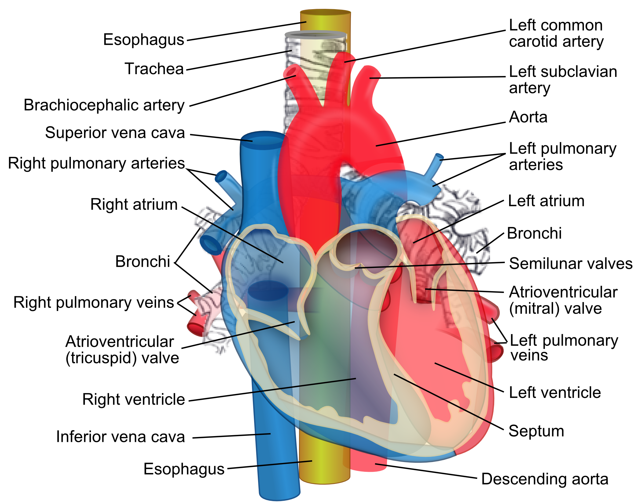
Chapter 19 Blood Vessels from image.slidesharecdn.com They let blood flow forward and prevent the backward flow. This article lists a series of labeled imaging anatomy cases by system and modality. Blood vessels comprise the vascular system. The 4 valves are the aortic, pulmonary, mitral, and tricuspid valves. Eventually, the smallest arteries, vessels called arterioles, further branch into tiny capillaries, where nutrients and wastes are exchanged, and then combine with other vessels that exit capillaries to form venules, small blood vessels that carry blood to a vein, a larger blood vessel that returns blood to the heart. It extends on each side of the neck and divides at the level of the larynx into two branches: Arteries (in red) are the blood vessels that deliver blood to the body. Use key choices to identify the blood vessel tunic described.
The width of blood vessels varies, but they all have a lumen.
The thick outermost layer of a vessel (tunica adventitia or tunica externa) is made of connective tissue. They can vary in size. The superior vena cava is the large vein that brings blood from the head and arms to the heart, and the inferior vena cava brings blood from the abdomen and legs into the heart. The venules and veins returning blood to the heart. The common cartoid artery extends from the brachiocephalic artery. The common carotid arteries have. It extends on each side of the neck and divides at the level of the larynx into two branches: Blood vessels form a continuous path for blood flow that starts and ends at the heart.arteries carry blood away from the heart, regardless of the degree of blood oxygenation.veins carry blood toward the heart. This layer is known as either tunica externa or advetitia. Blood vessels are the channels or conduits through which blood is distributed to body tissues. Blood vessels comprise the vascular system. Blood vessel labeling online quiz; Published by admin on june 18, 2020.
When the heart contracts, it pumps blood out through the arteries. To play this quiz, please finish editing it. They are designated as resistance vessels since they can regulate blood flow velocity by means of their respective muscle walls (approximately 120 mm hg). Vessel networks deliver blood to all tissues in a directed and regulated manner. They let blood flow forward and prevent the backward flow.
Blood Vessels Labeled Heart Anatomy Anatomy And Physiology Vessels Labeled Diagram Blood Vessels Labeling Exercises Cat Blood Vessels Labeled Human Anatomy Blood Vessels Human Blood Armando Steil from i0.wp.com Arteries and veins are composed of three tissue layers. •formed where capillaries unite • extremely porous 1) venules: The width of blood vessels varies, but they all have a lumen. Name the blood vessel labeled 'c'. Best quiz blood vessel labeling; The venules and veins returning blood to the heart. Bulky middle tunic contains smooth muscle and elastin 3. Blood vessels consist of arteries, arterioles, capillaries, venules, and veins.
Very small branches that collect the blood from the various organs and parts are called venules, and they unite to form veins, which return the blood to the heart.
Best quiz blood vessel labeling; Name the blood vessel labeled 'b'. Blood is supplied to the brain, face, and scalp via two major sets of vessels: All blood vessels consist of a similar basic structure, which includes: The middle layer is known as tunica media and is comprised of thin muscular tissue. The width of blood vessels varies, but they all have a lumen. Blood vessels are the channels or conduits through which blood is distributed to body tissues. The walls of blood vessels differ depending on the type of vessel. Ap2 learn with flashcards, games, and more — for free. The venules and veins returning blood to the heart. Blood vessels are the specially designed tubes that carry blood throughout the body. Arteries and veins are composed of three tissue layers. Classification & structure of blood vessels.
Arteries and veins are composed of three tissue layers. Name the blood vessel labeled 'd'. Name the blood vessel labeled 'b'. Ap2 learn with flashcards, games, and more — for free. This layer is known as either tunica externa or advetitia.
Great Vessels Wikipedia from upload.wikimedia.org Name the blood vessel labeled 'd'. This article lists a series of labeled imaging anatomy cases by system and modality. Normal function of the brain's control centers is dependent upon adequate supply of oxygen and nutrients through a dense network of blood vessels. There are five main types of blood vessels: The word vascular, meaning relating to the blood vessels, is derived from the latin vas, meaning vessel. Free online quiz blood vessel labeling They can vary in size. Vessel networks deliver blood to all tissues in a directed and regulated manner.
•formed where capillaries unite • extremely porous 1) venules:
The 4 valves are the aortic, pulmonary, mitral, and tricuspid valves. The outermost layer is comprised of connective tissue. Tunica intima—this is the inner thinnest layer. This set is often in folders with. Aside from capillaries, blood vessels are all made of three layers: The common cartoid artery extends from the brachiocephalic artery. They can vary in size. Blood pressure results from the blood flow force generated by the pumping heart and the resistance of the blood vessel walls. Blood vessels are the specially designed tubes that carry blood throughout the body. Arteries, arterioles, capillaries, venules and veins. Review the major systemic arteries of the body including those of the neck, arm, forearm, abdomen, pelvis, thigh, and leg in this interactive tutorial. Its smooth surface decreases resistance to blood flow Name the blood vessel labeled 'b'.